There was a time when every health issue women suffered was blamed on her hormones the haywire cycles, the lack of production (or more of it) and serious issues were related to her reproductive system cysts in the ovaries, blocked tubes, uterine fibroids these were the most common health concerns. There was very less knowledge of how the other major organs of her body like the heart, liver, kidneys were functioning or affected. A common myth that plaque the general public is that a woman s hormones protect her from organ failures. But in the modern times, we are seeing just the opposite. The number of women falling prey to diseases related to the major organs is just rising in numbers.
Dr. K. S. Shiva Kumar is one of the most reputed urology hospital in bangalore with many years of experience provides comprehensive urology treatment in bangalore.
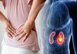
Kidneys are the organs that filter waste products from the blood. They are also involved in regulating blood pressure, electrolyte balance, and red blood cell production in the body. Many women are diagnosed with chronic kidney disease at a later stage of their lives, especially beyond 55 years of age. According to worldwide population-based data on the global burden of chronic kidney disease (CKD) in 2010, CKD is more prevalent in women (272 million) than in men (226 million). According to Global Prevalence of CKD A Systematic Review and Meta-Analysis CKD is more likely to develop in women compared with men, with an average 14 percent prevalence in women and 12 percent in men.
What makes women prone to kidney diseases?
Diabetes is one the biggest risk factors for chronic kidney disease. Blood pressure is second risk factor the third risk factor is NSAIDS. Apart from this, urinary tract infections (UTIs) are common risk factors for kidney disease more frequent in women than in men. HIV infection and complications of pregnancy, including pre-eclampsia, are also risk factors for CKD in females. The urethra is shorter in women than in men, which makes it easier for bacteria to travel from outside the body to the bladder. Once in the bladder, an infection can spread to the kidneys.
Yes, but there are few who might have more risk factors than the others. Kidney disease is considered as a risk factor for adverse pregnancy outcome and reduced fertility. Pregnant women are at even higher risk of a kidney infection. Some kidney diseases, such as lupus nephropathy or kidney infection (acute or chronic pyelonephritis), usually affect women more. Kidney infections (as most urinary tract infections) are more common in women and more likely to involve pregnant women.
What treatment is suggested for women who have kidney problems?
When kidneys fail, dialysis is required to remove wastes from the blood, however, it does not replace all of the functions of the kidneys, such as producing hormones. While all menopausal women are required to take calcium to prevent osteoporosis, it is important for those on dialysis. Because women on dialysis, their hormone levels might already be compromised. To help combat osteoporosis, additional calcium in the diet, or in the form of supplements, can help prevent bone loss.
How can women safe them from kidney diseases?
Many women with diabetic kidney disease may not have any symptoms. The only way is to get your kidneys checked is by blood (renal profile with electrolytes) and urine test. People with diabetes should be screened regularly for kidney disease at least once a year.
Making patients with Chronic Kidney Disease (CKD) drink more water does not slow down the decline of their kidney function, suggests a recent research.
“While many claims about the benefits of increased water intake remain untested, a growing body of evidence suggests that increased water intake improves kidney function through the suppression of the antidiuretic hormone.
SSurocare Hospital Bangalore is the best urology hospital in bangalore, with experienced top urologists, providing urology treatment & kidney specialist treatment.
This leads to the question of whether increased water intake can slow the progressive loss of kidney function.
Guided by a successful pilot study, the research team constructed a randomised clinical trial with patients at various hospitals. They wanted to determine if coaching patients to drink more water slowed their decline in kidney function over one year compared with those in the control group who were coached to maintain usual fluid intake.
“It is really about personalised medicine.
Could we look at the general population of patients with kidney disease and find that increased water intake helps slow the decline in function? And if not, how can we apply this knowledge to create more targeted treatment approaches?”
The trial was done on 630 participants with Stage 3 CKD. The cause of their disease was varied, including diabetes and high blood pressure.
“Those diagnosed with CKD have some degree of kidney damage, indicated by a decreased level of kidney function,” explained researcher.
“Kidney disease can range from mild to severe and, in some cases, leads to kidney failure. Kidney disease often starts slowly and develops without symptoms over a number of years, so it may not be detected until it has progressed to the point where kidney function is quite low,” urologist added.
Most patients with Stage 3 CKD experience either no symptoms or only mild effects, but they have already lost over 40 to 70 per cent of kidney function.
The research team found that after one year, the increase of water did not slow the loss of kidney function. They did find that an increase of water, particularly when the participant’s previous intake was low, did significantly suppress their antidiuretic hormone release. The prior benefits shown for water intake may relate to low water drinkers in the population.
Kidney stone disease is widespread in India, with an expectancy of 12 per cent in an overall population indicated to be prone to urinary stones.
Here At Shree Srinivasa Urocare, we have a team of highly experienced urologists. We provide best kidney stone treatment in bangalore at affordable cost.
Urolithiasis or nephrolithiasis refers to the formation of urinary calculi or kidney stones. This condition causes an incredible amount of pain in the abdomen and flanks. In developed countries, roughly 10-12 pr cent of the population is affected by kidney stones. Over the last few decades, drastic changes in diet and lifestyle have led to an increase in the severity of kidney stones disease (KDS) while the age of diagnosis has decreased. Men are twice as prone as women (6 per cent) to developing kidney stones. Also, it has been noticed that the first occurrence of stones typically appears between the age of 20 and 40. Around half of patients who have had kidney stones may develop another stone within 5 years. Today, as we celebrate National Nutrition Week, it is important to remember that this can be countered by following a proper nutrition.
KIDNEY STONES RAMPANT IN INDIA
In the Indian context, KSD is widespread, with an expectancy of 12 per cent in an overall population indicated to be prone to urinary stones. Out of this 12 per cent, 50 per cent are seriously impacted by renal damage, which often results in kidney loss. Since kidneys are vital life-sustaining organs, performing many functions to keep the blood clean and constantly maintaining the electrolytes and water content of the body, it is imperative to follow a proper nutrition-rich diet. Nutritional management of kidney stones aims to keep existing stones from growing larger and new stones from developing. While kidney stones are often treated with medication, changes in eating habits can mitigate the various complications.
NUTRITION CAN HELP REMOVE KIDNEY STONES
Here is a list of nutrition-rich items that can aid in removing kidney stones:
Drink plenty of liquids
Bypassing kidney stones can be extremely painful. Drinking plenty of fluids is the single most essential thing that individuals who suffer from kidney stones can act on to break down existing stones and avert future stones. As per a 2015 National Kidney Foundation meta-analysis, people who produced 2 to 2.5 litres of urine a day were 50 per cent less prone to developing kidney stones than those who produced less. Water is more effective than other fluids and it requires nearly 2 litres of water to enhance the function of the kidneys. Although untreated water has no nutrient benefit, it transports vital nutrients throughout the body. Other fluids which are beneficial include:
Orange, pomegranate and fresh tomato juice have been proven to be effective in preventing stone formation as they are rich sources of citrate and magnesium.
Lemonjuice can increase urine citrate and likely reduce kidney stone risk.
Watermelon juice is diuretic and high in potassium salts, which aid in the regulation of urine acidity and the removal of small stones and crystals.
Apple Cider Vinegar contains citric acid, which facilitates the dissolution of kidney stones by alkalizing the blood and urine, making it easier to flush out toxins in the kidney.
Increase your intake of calcium-rich foods
Since dietary calcium binds oxalate in the intestines, less is absorbed by kidneys and lower concentrations end up in urine. Yoghurt, lentils, seeds, soy products, and beans are all excellent sources of calcium-rich food. Kidney beans, for instance, have a positive impact on health when eaten as a substitute for meat or other high-cholesterol protein sources. Kidney beans resemble the shape of a kidney and the Vitamin B present in them, aids in the flushing out of kidney stones. Kidney beans are also a good source of fiber and contain a lot of minerals, which enable better urinary tract function.
Choose magnesium-rich vegetables and wholegrain foods
Leafy greens are super healthy, and many are loaded with magnesium, which boosts kidney function and helps to dissolve stones. Kale, spinach, collard greens, turnip greens, and mustard greens are among the vegetables high in magnesium. Magnesium reduces oxalate absorption, so people who consume more magnesium reduce their risk of developing kidney stones. A high-fiber diet and choosing whole-grain foods can also lower the risk of kidney stones growing bigger by reducing calcium absorption and oxalate absorption.
TAKEAWAY
Maintaining a nutritionally dense diet is beneficial for kidney function, but breaking certain habits is even more important. Here are some pointers to remember:
If you are overweight or obese, make an effort to lose weight.
Limit high purine foods.
Reduce the amount of sugar and salt in your food and beverages.
Kidney stones are solids formed in the kidneys when substances like calcium, oxalate and phosphorus that are excreted through the urine become concentrated. A kidney stone may either settle down in the kidneys or travel through the urinary tract. Kidney stones usually vary in size. So a smaller crystal may comfortably travel down the urinary tract and get expelled on its own without causing any pain or discomfort.
However, when the crystals increase in size they may from larger hard stones which tend to get stuck as they pass along the urinary tract. This can cause several problems. Mainly it may block the usual flow of urine, causing extreme pain and bleeding. Other symptoms of kidney stones include pain in the lowerback region, vomiting and nausea.
Various factors put an individual to risk for kidney stones. Most common factors include a family history of kidney stones, repeated urinary tract infections, blocked urinary tract, or any health condition that increases the level of substances usually excreted through the urine. Lack of sufficient intake of water and certain medications can also cause kidney stones. Prevention of kidney stones include medications and dietary modification.
Types
There are four types of kidney stones which include:
Uric acid stones: The factors that can contribute to the formation of uric acid stones is a high protein diet, malabsorption, diabetes, chronic diarrhoea or metabolic syndrome.
Calcium stones: Calcium stones are the most prevalent type of kidney stones. These are in the form of calcium oxalate stones.
Metabolic disorders, high consumption of vitamin D can increase oxalate levels in urine. Another form in which calcium stones are found is calcium phosphate stones. Medical treatment of migraines with topiramate may also be associated with the formation of calcium stones.
Cystine stones: The aetiology of cystine stones is related to cystinuria, a hereditary condition that promotes kidneys to excrete a higher amount of amino acid.
Struvite stones– these stones are caused due to a urinary tract infection. Struvite stones are large and fast-growing.
Symptoms
Unfortunately, kidney stones do not cause any symptoms until they move around in your kidney or pass through your ureter, the tube connecting the kidneys and urinary bladder. Small stones may pass out even without causing any symptoms. But, with larger stones, the following symptoms may be seen:
Back and groin pain
Pain on urination
Abnormal urine colour
Nausea and vomiting
Persistent urge to urinate
Causes And Risk Factors
Causes
Accumulation of uric acid, calcium or oxalate in the urine in the levels higher than your urine can dilute, can cause Kidney stones. These accumulations are in the form of crystals.
Risk Factors
As men have more muscle mass than women, they are at an increased risk of kidney stones. This is because the daily breakdown of muscle mass leads to a tendency to form stones in the kidneys.
Other common risk factors of kidney stones include –
Family history: You are more susceptible to develop kidney stones if someone in your family has had stones.
Personal history: The risk of having a kidney stone is much greater if you already have a stone.
Age: Though kidney stones can occur at any age, they are most commonly diagnosed in adults aged 40 and above.
Obesity: High body mass index (BMI), large waist size and weight gain have been linked to an increased risk of kidney stones.
Dehydration: Less fluid intake will increase your risk of getting kidney stones. People that live in hot regions or those who work in environments where they tend to sweat a lot will have a higher risk of developing stones.
Existing medical conditions: Gastric bypass surgery, inflammatory bowel disease or chronic diarrhoea may cause changes in the digestive process that affect your absorption of calcium and water, increasing the levels of stone-forming substances in your urine. Renal tubular acidosis, cystinuria, thyroid problem (hyperparathyroidism) and urinary tract infections can increase stone formation.
Certain diets: Eating a diet that’s high in animal protein, sodium and has low fibre content can increase your risk for kidney stones. This is especially true for a high-sodium diet. Too much sodium increases the calcium load on your kidneys, significantly increases your risk of kidney stones.
Prevention
Here are 10 simple ways to prevent kidney stones –
Drink lots of water
Drink healthy beverages
Eat smartly
Eat more dairy foods every day(no scientific data)
Limit the intake of seafood
Reduce the intake of salt
Maintain a healthy weight
Know your risk
Don’t hold your pee for too long
Avoid alcohol and cigarettes
Diagnosis
Ultrasound of the kidney, ureter and bladder (KUB) is the most convenient and frequently used investigation to diagnose kidney stones. An X-ray KUB can further assist in diagnosis. However, if the stones are small, the patient is obese, or there is an unusual formation of gas in the abdomen, a CT scan is a better method of diagnosis.
Urine testing is significant in the detection of stone-forming minerals. A blood test can help in marking high levels of calcium or uric acid, which help in the diagnosis of kidney stones.
Treatment
Treatment is based on the size of the stones. Smaller stones can be treated with plenty of fluids, pain medications and certain antispasmodic drugs (pain and spasm relieving), which help in passing the stone out. But even when the symptoms disappear, it is essential to confirm whether the stone has been eliminated through an ultrasound or a CT scan.
Larger stones which do not pass out easily require medical therapy and more advanced treatments such as –
Sound waves: Depending on the size and location of the stones, extracorporeal shock wave lithotripsy (ESWL) may be done. ESWL uses sound waves to create strong vibrations (shock waves) that break the stones into tiny pieces, making them easier to be passed through urine. The procedure lasts for about 45 to 60 minutes. It can cause moderate pain, so it is preferably performed under sedation or light anaesthesia. ESWL can also cause blood in the urine and discomfort as the stone fragments pass through the urinary tract. Depending on the size of the stones, one or more sessions may be needed.
Percutaneous Nephrolithotomy (PCNL): This treatment is used for very large stones or where ESWL is unsuccessful. The procedure involves surgically removing the stone using small telescopes and instruments inserted through a small incision in the back.
Ureteroscopic Removal: Smaller stones in the ureter or kidneys may be removed by passing a thin, lighted tube (ureteroscope) equipped with a camera through the urethra and urinary bladder to the ureter. Once the stone is located, special tools can snare the stone or break it into pieces, making them easier to pass through the urine. A small tube (stent) may be placed in the ureter to relieve symptoms like swelling and promote healing.
Laser Lithotripsy: Some stones cannot be treated with ESWL because of their size, location, type. In such cases, ureteroscopic laser lithotripsy may be used. In this procedure, a tiny fiberoptic camera is passed into the urinary tract through the urethra to locate the stones in the bladder, kidney or ureter. Then using laser technique, larger stones are fragmented into thousands of small pieces, which are flushed out through urine.
Lifestyle/management
Stay hydrated – a daily requirement of drinking enough fluids should be fulfilled to pass about 2 litres of urine all day.
Salt restriction – restrict the amount of salt in your diet.
Avoid oxalate-rich food, such as spinach, beets, okra, tea, black pepper, Soy products and chocolate should be restricted as they are found to have higher levels of oxalate.
Choose calcium-rich foods instead of calcium supplements – calcium-rich food does not affect kidney stones; however, calcium supplements increase the risk of kidney stones. Therefore, use calcium supplements with caution.
Prognosis And Complications
Prognosis
The chances of recurrence of kidney stones are quite high. More than half of the people, who have had a stone in the past, will develop a new stone in their lifetime.
Complications
Untreated kidney stones can significantly damage the kidneys and even lead to end-stage renal disease (ESRD), requiring dialysis. But early detection and treatment may completely reverse the damage, even in those on dialysis.
Unfortunately, many a time, kidney stones remain undetected for years because minor symptoms are ignored/neglected. By the time the patient consults a doctor, the damage becomes irreversible or only partially reversible.
Women of all ages suffer from various types of bladder problems, which vary as to cause and contributing factors. Some are due to weakness of the pelvic fascio-muscular supports, while in other women the problem is caused by nerve damage. In some women, the bladder control is affected by various medications for neurologic problems.
The urologist in bangalore, offers on-site diagnostic testing services to evaluate the health of your pelvic floor organs. We also offer customized treatment plans to prevent urine leaks from affecting your quality of life.
To help women with regaining bladder control, it is important to identify the type of bladder control that is affected in each case. The main types of bladder problems in women are temporary, stress, urge, and mixed incontinence, but other pathologies may be observed as well.
Temporary or transient incontinence
Temporary or transient incontinence is a short-lived episode of loss of control over the escape of urine from the bladder. The condition is present in practically the third of community-dwelling elderly individuals, and in more than half of hospitalized patients.
A urinary infection may stimulate bladder activity leading to incontinence. Some medications lower the cortical control over the urinary bladder or cause overproduction of urine. Pregnancy is another cause of temporary incontinence. In all these cases, continence is restored when the etiologic factors are found and corrected.
Stress incontinence
In this situation, the woman reports that urine escapes when she coughs, laughs, sneezes or does vigorous exercise – anything which puts repeated stress on the pelvic floor. This type is most common among women.
In a strong pelvic floor, the muscles contract on reflex as soon as there is an increase in intra-abdominal pressure. Their contraction shuts off the urethra, preventing the escape of urine. However, when these supports are weakened, any sudden rise in pressure allows urine to flow out through an open urethra.
Thus pregnancy or childbirth changes, as well as the thinning that occurs with menopause, may all precipitate stress incontinence. Estrogen drops in the specific phases of the menstrual cycle may weaken the urethra and also provoke stress incontinence.
Urge incontinence
Some women report that when they feel the need to pass urine, the urge is strong, sudden and uncontrollable, being followed by the escape of urine almost at once. This points to the loss of bladder control from nerve damage, as occurs in diabetes mellitus, a cerebrovascular accident, or an infection.
Mixed incontinence
Mixed incontinence represents a combination of urine leakage following a sudden sneeze or cough, along with other episodes of urge incontinence. It is also considered the leading cause of incontinence in women in the community, but also in the hospital setting.
Functional incontinence
Functional incontinence is not so much a problem of the bladder as it is of the affected person’s mobility and cognition. If, for example, a woman cannot move to the bathroom whenever she needs to, she may urinate where she is once the bladder capacity is far exceeded. Furthermore, the ability to urinate on cue may be affected by physical or mental weakness.
Overactive bladder
Women who complain of having to pass urine eight or more times a day, or of waking up two or more times to visit the bathroom every night, may have an overactive bladder. Since the detrusor muscle contracts unpredictably, these patients may also have urge incontinence.
Overflow incontinence
Overflow incontinence refers to the type of urinary leakage that occurs because the bladder does not empty completely during urination. Thus the bladder has a significant amount of residual urine, and overfills rapidly, leading to overflow of urine within a short time. It is often encountered in conditions such as diabetes and multiple sclerosis.
The changes your body goes through during pregnancy can be both wondrous and worrisome. If you’re experiencing unexpected urine leaks during or after pregnancy, don’t panic. There are things you can do to restore your bladder control and your self-confidence.
The urologist in bangalore, offers on-site diagnostic testing services to evaluate the health of your pelvic floor organs. We also offer customized treatment plans to prevent urine leaks from affecting your quality of life.
Understanding urinary incontinence Urinary incontinence is the inability to control urine flow. Typically, your bladder holds urine until it’s ready to leave your body through the urethra. As your bladder fills with urine, the bladder muscles relax to create space. Bladder sphincter muscles hold your bladder closed until you go to the bathroom.
When you have urinary incontinence, this process doesn’t work properly. You might experience urinary incontinence because of a pregnancy or childbirth. Other common causes of urinary incontinence in women include:
Obesity
Constipation
Menopause
Neurological disorders
Urinary tract infections (UTI)
You can also develop urinary incontinence if you have tumors or other obstructions in your urinary tract. For this reason, you should seek a diagnostic evaluation at the Urology Specialist Group to prevent more serious complications.
Types of urinary incontinence There are four types of urinary incontinence that our urologists can diagnose and treat.
Stress incontinence Stress incontinence develops when too much pressure is placed on your bladder, such as during a pregnancy.
Urge incontinence Urge incontinence causes a sudden and urgent need to urinate. You might not make it to the bathroom on time before urine starts leaking out.
Overflow incontinence Overflow incontinence occurs when you can’t fully empty your bladder when using the bathroom, allowing the leftover urine to leak out.
Functional incontinence Functional incontinence describes a loss of bladder control that occurs because of physical or mental complications.
How pregnancy plays a role in urinary incontinence Urinary incontinence is common in pregnancy because your baby grows a little every day and puts pressure on your bladder. This is especially true in your final weeks of pregnancy when even small sneezes and coughs can cause urine to leak out.
Childbirth is another factor that contributes to urinary incontinence. When you give birth, your pelvic floor muscles stretch out and weaken.
Since the pelvic floor muscles are responsible for keeping your bladder in place, you may experience more frequent urine leaks when they can no longer support your bladder or other pelvic floor organs.
Women are also at risk for pelvic organ prolapse, a condition that affects your bladder and other pelvic floor organs. When your pelvic muscles are too weak to support the organs, they can fall down into your vaginal canal and even protrude from your vagina.
Treatments for urinary incontinence If you have urinary incontinence due to a pregnancy or childbirth, our urologists can design a treatment plan to reduce urine leaks and prevent prolapse and other complications.
To start, you may need to make lifestyle and diet changes to stop urine leaks. These changes can include:
Avoiding caffeine and other stimulants
Emptying your bladder on a set schedule
Doing pelvic floor exercises
Maintaining a healthy body weight
We also offer pelvic floor reconstruction surgery to repair the muscles that keep your bladder in place. During this type of surgery, our surgeons put your pelvic organs back into their original position and tighten your pelvic floor muscles to support the organs.
If you’re embarrassed by frequent or unexpected urine leaks, visit our Urology Hospital in Bangalore near you today or schedule a diagnostic evaluation online.
Many medical conditions are straightforward and easy to explain. However, some occur and cause a person to just not feel right – like something is out of place. For women, this could be a sign of pelvic floor prolapse.
Here at SSurocare we are best urologist in bangalore, we provide the best solutions for Urinary problems.
Many women (about 30-40%) experience pelvic floor prolapse or “dropped bladder” during their lifetime. Pelvic floor prolapse can cause a sensation that tissues or structures in the vagina are out of place. The condition occurs when the weight-bearing or stabilizing structures that keep the vagina in place weaken or deteriorate. In turn, the upper portion of the vagina loses its normal shape and sags or drops down into the vaginal canal or even outside of the vaginal opening.
A pelvic floor prolapse can be caused by childbirth, menopause, a hysterectomy, obesity and advanced age. When pelvic floor prolapse occurs, a variety of symptoms can appear depending on the type of prolapse. Click here to learn about the four types.
Symptoms may include:
Pressure or fullness in the vagina or pelvis Painful intercourse (dyspareunia) Recurrent urinary tract infections (UTIs) Difficulty emptying the bowel and/or bladder Constipation Urinary stress incontinence Pain that increases during long periods of standing A lump or protrusion of tissue at the opening of the vagina Symptoms don’t always appear, so some women may have pelvic floor prolapse and not know it. However, if symptoms do surface, solutions are available. Treatment options depend on the severity of the prolapse, whether the woman is sexually active, and her personal preferences. In some cases, lifestyle changes or exercise may suffice.
Nonsurgical treatments include:
Activity modification, such as avoiding heavy lifting or straining Kegel exercises, which are simple strengthening exercises that can tighten the muscles of the pelvic floor A pessary (a small plastic or silicone medical device) can be inserted into the vagina for support Estrogen replacement therapy can help strengthen and maintain muscles in the vagina In some situations, surgery may be required as most worsening pelvic floor prolapses can only be corrected with surgery. A variety of surgical treatment options are available, depending on the woman’s situation and preferences.
If you are experiencing symptoms of a pelvic floor prolapse, don’t ignore discomfort or the feeling that something is out of place. A urologist can provide the diagnosis and recommendations for proper treatment.
Drinking plenty of water each day may have an unexpected benefit — staving off urinary tract infections, a new study reports.
You can contact our best Urologist in Bangalore for treating both kidney stones just as back torment, notwithstanding, it is critical to perceive the side effects and pick the correct specialist appropriately.
Young women plagued by UTIs who drank an additional 6 cups of water each day were nearly half — 48 percent — as likely as a control group to have another infection, the study showed.
The water group also reduced their use of antibiotics by roughly half — or 47 percent.
What’s more, the amount of time that passed before a woman’s next UTI and between recurrent infections was longer in the water group than in the control group.
Women are more prone to UTIs than men, partly due to their anatomy. A shorter urethra makes it easier for bacteria from rectum to enter the vagina and travel to the bladder.
“Flushing bacteria out of the bladder, it’s been known, at least it’s been thought, that it is protective. This study suggests that it is,”.
“It is the urine output which is of course the key factor leading to the effect seen in the study,”
She cited a 2010 review estimating that 11 million to 12 million women a year will suffer from a UTI, accounting for 1.6 billion antibiotic prescriptions a year.
“We might not even need to use antibiotics,” he said.
The study, conducted in Bulgaria, involved 140 healthy premenopausal women who had at least three UTIs in the past year. All reported low daily fluid intake. They drank roughly four cups of fluid per day, including just 2 cups of water a day.
Half of the women in the study were asked to consume 1.5 liters of water, or about 6 cups, each day in addition to their usual daily fluid intake. Specifically, they were asked to begin drinking a 1/2-liter water bottle at each meal and finish each bottle before the next meal.
The remaining women served as a control group. They continued their usual fluid intake.
The women were followed for a year, with clinic visits at the beginning of the study as well as 6 and 12 months later. Researchers measured their water and fluid intake; urine volume, frequency and concentration; and symptoms. They also called the women each month to assess compliance and counsel them to return to the clinic for evaluation and possible treatment if they were experiencing UTI symptoms.
Overall, the water group increased daily water intake by close to 5 cups a day. Their total daily fluid intake, including water and other beverages, averaged nearly 12 cups.
By contrast, the control group’s total daily fluid intake was less than half of that.
Hooton noted that it’s not clear exactly how much fluid intake must increase to deliver a benefit.
“There’s no magic in a liter-and-a-half,” he said.
Likewise, he believes any increase in fluid — not just water — may be beneficial since fluids are mostly water. And while the study involved younger women, he said older women who experience recurrent UTIs may benefit from increasing their daily fluid intake as well.
Whether the advice would benefit women with normal urine output, however, is unclear, Wessells noted.
“In addition, the bother associated with the higher urine output, and feasibility of implementing this water intake strategy across a range of occupations and ages, requires further study as well,” he said.
The urinary system is comprised of the kidneys, ureters, bladder, and the urethra. The purpose of the urinary system (also called the renal system) is to remove waste from the body and to regulate electrolytes, blood volume, blood pressure, and blood pH. The kidneys filter the blood so the waste can exit through the ureters to the bladder in the form of urine.
If you have any questions regarding urology problems contact us shree srinivas uro care urology hospital in bangalore. You can also get in touch with us through our social media channels such as facebook and youtube.
Diseases that affect the urinary system are grouped under an umbrella term of urologic diseases. These diseases can arise from a genetic defect or an acquired illness. The illness may arise from the urinary system itself, or it could be secondary to a disease of another bodily system. For example, in diabetes, the kidneys become vulnerable to hypertension.
Kidney Disease
The kidneys represent a central organ of the urinary system. Disease of the kidney can lead to renal failure, which is a serious, life-threatening condition, and the etiology can be divided into pre-renal, intrinsic and post-renal failure.
Renal failure that occurs as a result of impairment of the blood supply to the functional kidney cells (nephrons) is known as pre-renal failure. Diseases causing pre-renal failure include renal artery stenosis, intravascular volume depletion, relative hypotension, compromised cardiac output, as well as hepatorenal syndrome.
Intrinsic renal failure is caused by diseases of the functional tissue of the kidney, or the parenchyma. Forty-five percent of intrinsic acute renal failure is caused by acute tubular necrosis.
Post-renal acute kidney failure is caused by an obstruction of the urinary tract by an object such as a clot, a kidney stone, or a tumor. Almost twenty percent of community-acquired renal failure can be a result of some process that results in post-renal etiology of kidney disease.
Urologic diseases affecting parts of the urinary system other than the kidneys include urinary tract infections, benign prostatic hyperplasia, obstructions of the urinary ducts, and malignancies of various localizations.
Diagnosis
Blood tests for urea and serum creatinine can indicate the functional status of the kidneys, potentially identifying the presence of a renal disease. Urinalysis is used to check for infection or high levels of protein indicating a problem. The flow of urine from the bladder can be measured by urodynamic testing, while ultrasound is used for imaging studies of the kidneys and the bladder.
Treatment
Urologic diseases have many potential treatments, some of them making use of newer technologies. Minimally invasive surgeries are now available for kidney disorders, prostate, and reproductive systems – including laser prostatectomy.
Microwave therapy is a procedure used for treatment of disorders of the lower urinary tract caused by benign prostatic hypertrophy. It is carried out by insertion of a catheter into the bladder to position a microwave antenna near the prostate gland. The microwave antenna then safely heats and destroys prostate tissue.
Another newer treatment that is available in the area of urologic disease is laser therapy for kidney stones. Kidney stones are formed from crystals of calcium, ammonia, urea, and other substances that build up over time. They are most common in men between 30 and 60 years of age. The laser is delivered by a fine wire, guided by a tiny camera that can reach the stones without invasive surgery and break them apart, so that they can pass out of the body.
Cholesterolosis is a common affection of the biliary system, characterized by the accumulation and deposition of cholesterol inside the gall bladder and in its mucosal membranes. Cholesterolosis usually occurs due to chemical imbalances at the level of the biliary system and the disorder is rarely associated with high serum cholesterol levels, diabetes mellitus or atherosclerosis. Cholesterolosis generally occurs on the premises of inappropriate activity of the gall bladder and changes in the composition of bile, facilitating the deposition of cholesterol inside the gall bladder and biliary ducts. In the absence of an appropriate treatment, cholesterolosis can lead to serious complications, including gall bladder cancer.
If you have any questions regarding urology problems contact us shree srinivas uro care urology hospital in bangalore. You can also get in touch with us through our social media channels such as facebook and youtube.
Although this type of gall bladder disorder can occur in both sexes, cholesterolosis commonly affects women. Also, cholesterolosis has the highest incidence in people with ages over 50. Most patients with cholesterolosis are asymptomatic, rarely experiencing symptoms such as diffuse abdominal pain or discomfort. While the disorder is treatable in its early stages, advanced forms of cholesterolosis require surgical intervention. Medical reports indicate that cholesterolosis is responsible for more than 50 percent of cholecystectomies (surgical procedure that involves removal of the diseased gall bladder).
Cholesterolosis can affect the gall bladder locally or generally. General forms of cholesterolosis appear as inflammation of the gall bladder mucosa, corroborated with yellow staining of the gall bladder tissues and membranes (due to deposition of fat). Localized forms of cholesterolosis are characterized by the formation of small polyps, soft prominences that emerge from the gall bladder interior walls. The size of these polyps varies from 1 to 10 mm.
Cholesterolosis can be only be revealed by modern scanning techniques such as ultrasound imaging. Ultrasound tests can quickly unveil the presence of polyps and lipidic masses associated with cholesterolosis. Polyps appear as immobile prominences attached to gall bladder mucosal walls. The presence of these prominences rarely involves hardening or thickening of the gall bladder interior membranes. Patients who present smaller polyps usually receive medication treatments for overcoming the disorder. However, the presence of larger polyps often involves cholecystectomy. Patients confronted with such gall bladder problems may also receive biopsies before surgery. Although gall bladder polyps are usually benign, cholesterolosis can also lead to malignant activity at the level of the biliary system.
Similar to cholesterolosis, adenomyomatosis is a disorder that can also lead to malignant cellular activity at the level of the gall bladder. Unlike most forms of cholesterolosis, adenomyomatosis is characterized by thickening of the gall bladder mucosal walls. In order to distinguish between the two disorders, doctors commonly inspect the integrity and the general aspect of the gall bladder walls before deciding upon the final diagnosis. Speed is vital in diagnosing and treating gall bladder disorders such as cholesterolosis and adenomyomatosis, as both these affections can lead to malignancies. Prompt medical intervention can easily make the difference between complete recovery and partial recovery that exposes patients to a high risk of malignant disease.